
Our Services
Digital X-Rays
Digital X-rays in Royal Oak, Michigan, help reveal areas of your mouth that are not visible during a visual exam, guiding accurate diagnoses and timely care. At Royal Oak Smiles, this modern imaging supports preventive dentistry and treatment planning with fast, detailed images and very low radiation exposure.
Digital X-Rays Explained
Digital X-rays use electronic sensors to capture images of your teeth, roots, and jaw. Instead of film that must be developed, images appear instantly on a computer screen. This allows the dental team to adjust brightness and contrast, zoom in on details, and share findings with you chairside for clear understanding.
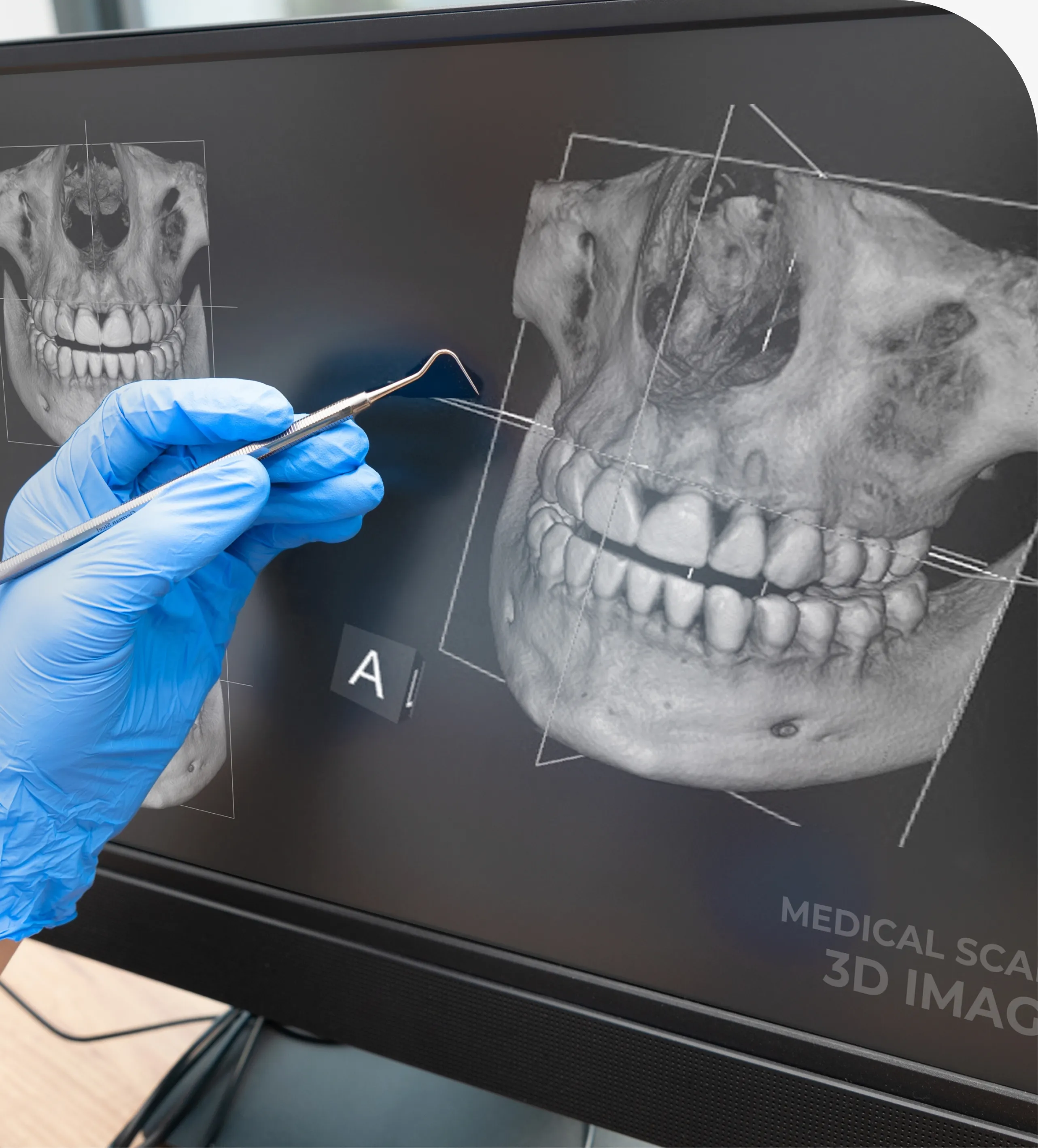
Common types include bitewing X-rays that show cavities between back teeth, periapical images that focus on a specific tooth from crown to root, and panoramic scans that capture the entire mouth in one image. Some offices may also use cone beam CT for complex cases such as implant planning; availability varies by practice.
Benefits of Digital Dental Radiography
- Lower radiation dose compared with traditional film systems.
- Instant images that speed visits and reduce retakes.
- High-resolution detail that helps spot decay, infection, and bone loss early.
- Easier communication as you can view images and discuss options together.
- Eco-friendly approach with no chemical processing.
- Secure storage for comparisons over time to track changes.
How Digital X-Rays Work
The process is straightforward and comfortable. A small sensor is placed where the image is needed, and a brief exposure captures the picture. For panoramic images, you stand or sit while the machine moves around your head to record the entire jaw. Images appear within seconds, so adjustments or additional angles can be taken right away if needed.
Digital systems follow the ALARA principle—“as low as reasonably achievable”—to limit radiation. A lead apron and, when appropriate, a thyroid collar provide extra shielding.
Your Digital X-Ray Experience
Most visits begin with questions about your oral health and risk factors. The team selects the type of images based on your needs—such as bitewings for cavity screening or periapicals to evaluate a painful tooth. The sensor is positioned gently, and each exposure lasts less than a second. You will breathe normally and remain still during the quick capture.
When Are Digital X-Rays Recommended?
- New patient exams to establish a baseline of your teeth and bone.
- Routine checkups to detect early decay or gum disease changes.
- Monitoring dental restorations, root canals, and implants.
- Assessing symptoms such as pain, swelling, or trauma.
- Planning treatments like extractions, orthodontics, or implants.
Frequency depends on your cavity risk, gum health, and history. Many adults with low risk may need bitewings every 12–24 months, while higher-risk patients and children may benefit from more frequent imaging. Our dentist will recommend the schedule that fits your situation.
What to Expect After Imaging
Once images are taken, our dentist will review them with you to explain findings, such as early decay between teeth or signs of bone loss from periodontal disease. These results help answer common questions like what digital dental X-rays show, how digital X-rays work, and the benefits of digital radiography in guiding care. If treatment is needed, images inform the plan and help you understand your options.
Frequently Asked Questions
Yes. Digital systems use very low radiation, far below everyday background exposure. Shielding, targeted beams, and the ALARA approach further limit dose.
It varies by risk. Low-risk adults may need bitewings every 12–24 months, while new patients, children, or those with active disease may need them more often.
Bitewing images check for decay between back teeth, periapicals examine a single tooth from crown to root, and panoramic scans view the entire mouth and jaws.
Yes, but X-Rays provide important information that a visual exam cannot. Discuss concerns so our dentist can explain risks, benefits, and possible alternatives.
Non-urgent images are often postponed. If necessary for urgent care, modern shielding and focused beams make dental X-Rays safe. Inform your dental team.
No. You may feel gentle pressure from the sensor. If you have a strong gag reflex, tell the team so positioning can be adjusted for comfort.
Ready to Get Started?
Schedule Your Visit Today
Contact Royal Oak Smiles to learn more or book a consultation with our experienced team.